Diabetic Retinopathy/Vein Occlusions/Retinal Tears
Our PASCAL laser system is the newest addition to our treatment modalities. This is one of the newest pattern lasers able to deliver laser treatment quickly with much less discomfort than previous laser systems. The PASCAL laser system often means fewer visits are required to obtain a full treatment than with regular laser systems. This means an overall better experience by our patients with less time spent obtaining treatment.Age Related Macular Degeneration
At Tulsa Retina Consultants we provide the full spectrum of treatment options for our patients with macular degeneration. This includes laser treatment, PDT, and intravitreal injections. Most often the best treatment for wet macular degeneration involves the delivery of a specialized drug into the vitreous cavity of the eye (intravitreal injection). This allows for more direct treatment to the problematic area. Three drugs are primarily used for the purpose: Eylea, Lucentis, and Avastin. These medications will be discussed with you as well as the risks and benefits of treating your condition.
Retinal Surgery
At Tulsa Retina Consultants we operate using the latest advancements in surgical equipment and technique. Sutureless vitrectomy has become the standard of care throughout the retina community. This means shorter surgical times, faster healing, and less inflammation without any increased risk to our patients. We desire to help you you recover your vision as quickly as possible and get back to your normal daily activities.

Imaging Modalities
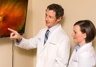
At Tulsa Retina Consultants we incorporate a wide array of imaging modalities to fully assist us in the diagnosis and management of your disease. These include:
- Optical Coherence Tomography (OCT) – a non-invasive, non-contact device that obtains high-resolution, cross-sectional image of the retina. This aids in the diagnosis and treatment of patients with macular diseases such as macular degeneration, macular holes, epiretinal membranes, diabetic macular edema and other macular diseases.
- Diagnostic Ultrasound – uses sound waves to form an image of the eye and is used to examine internal aspects of the eye.
- Fluorescein Angiography – involves the injection of a small amount of fluorescein dye (not Iodine dye) through a patient’s peripheral vein — usually the arm or hand. Shortly after, an ophthalmic photographer takes a series of retinal photographs. The fluorescein dye is then evaluated to determine areas of concern.
- Fundus Photography – a non-invasive diagnostic procedure that provides photographs of the back of the eye to help determine the health of retina and related structures.
- Visual Field Testing – a non-invasive, computerized, diagnostic procedure that evaluates your peripheral vision and provides information about the neurological function of the retina, optic nerve and brain.